3D imaging helps surgeons measure size of tumours
26 November/ updated 3 December 2014
Three-dimensional ultrasound imaging and augmented-reality software allows surgeons to pinpoint a tumour and measure its volume, including its depth, according to a study presented at the 2014 American College of Surgeons Clinical Congress. See video below.
Surgical oncologists, or cancer surgeons, usually remove breast cancers by relying on tactile feedback and radiologic images of the tumour, such as mammograms and ultrasound images. M. Catherine Lee, MD, FACS, coauthor of the study and associate professor of surgery at H. Lee Moffitt Cancer Center, Tampa, Florida, said, “Our goal in a lumpectomy is to get the lump out with a small rim of normal tissue around it, but we sometimes find out we did not get all of the tumour.”
Dr Lee’s team developed an imaging guidance system designed to minimize the need for repeated operations while sparing greater amounts of healthy breast tissue. Collaborating with St. Thomas University, Florida, the researchers designed a software algorithm that works with digital ultrasound technology. Ultrasound images are converted into 3D images on a computer screen.
In a simulated surgical procedure, the investigators studied the use of augmented-reality to fuse real-world and virtual 3D images. These augmented-reality images can then be transmitted to high-definition 3D glasses or other devices. When the surgeon wears such glasses, they see a superimposed, reconstructed 3D digital image over the actual tumour.
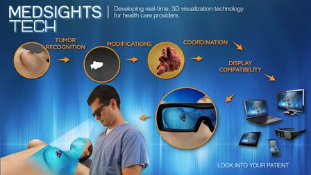
“It gives the impression of X-ray vision. You can see the tumour through the skin,” said lead author Segundo J. Gonzalez, MD, a surgical oncology fellow at Moffitt Cancer Center.
He and his co-investigators analyzed 66 ultrasound images of tumours (32 pictures of a single tumour and 34 of multiple tumours) inside a plastic model of a breast to determine the augmented-reality system’s accuracy of measuring tumour volume. The closer the 3D ultrasound image overlapped with the actual tumour, the more accurate the software was.
For detecting a single tumour, the volumetric accuracy was 1.2 cubic mm, which Dr. Gonzalez called “extremely accurate.” Likewise, accuracy for multitumour detection was 5.4 cubic millimeters, or 0.0003 cubic inches.
The investigators hope to study their software using a smart phone camera and, eventually, study it in patients. Dr. Gonzalez is commercializing the new technology through a start-up company, MedSights Tech Corp., in Tampa.
An animated illustration of the technology