Raman laser highlights tumour cells in brain tissue during surgery
10 September 2013
A new laser-based technology can identify cancerous cells during surgery, enabling surgeons to make sure they remove all the cells that could develop into a new tumour.
In a paper published in the journal Science Translational Medicine, a team of researchers from the University of Michigan Medical School and Harvard University describes how the technique allows them to identify the tiniest areas of tumour cells in brain tissue.
The basic technique used, stimulated Raman scattering (SRS), is named after CV Raman, one of the Indian scientists who co-discovered the effect and shared a 1930 Nobel Prize in physics for it. SRS can detect a weak light signal that comes out of a material after it’s hit with light from a non-invasive laser. The spectrum of light that is emitted by the sample gives details of its chemical makeup.
Over the past 15 years, Dr Sunney Xie of the Department of Chemistry and Chemical Biology at Harvard University has advanced the technique for high-speed chemical imaging. By amplifying the weak Raman signal by more than 10,000 times, it is now possible to make multicolour SRS images of living tissue or other materials. The team can even make 30 new images every second, the rate needed to create videos of the tissue in real time.
The team used this technique to distinguish tumour from healthy tissue in the brains of living mice and then showed that the same was possible in tissue removed from a patient with glioblastoma multiforme, one of the most deadly brain tumours. Now, the team is working to develop the approach, called SRS microscopy, for use during surgery to guide them in removing tissue, and test it in a clinical trial.
The need for a better solution
On average, patients diagnosed with glioblastoma multiforme live only 18 months after diagnosis. Surgery is one of the most effective treatments for such tumours, but less than a quarter of patients’ operations achieve the best possible results.
“Though brain tumour surgery has advanced in many ways, survival for many patients is still poor, in part because surgeons can’t be sure that they’ve removed all tumour tissue before the operation is over,” says co-lead author Daniel Orringer MD, a lecturer in the University of Michigan Department of Neurosurgery. “We need better tools for visualizing tumour during surgery, and SRS microscopy is highly promising,” he continues. “With SRS we can see something that’s invisible through conventional surgical microscopy.”
Highlighting the tumour
The new paper describes the first time SRS microscopy has been used in a living organism to see the “margin” of a tumour — the boundary area where tumour cells infiltrate among normal cells. That’s the hardest area for a surgeon to operate, especially when a tumour has invaded a region with an important function.
As the image shows, the technique can distinguish brain tumour from normal tissue with remarkable accuracy, by detecting the difference between the signal given off by the dense cellular structure of tumour tissue, and the normal healthy grey and white matter.
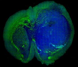
An image of a human glioblastoma brain tumour
in the brain
of a mouse made with SRS microscopy. The tumor
(blue)
is easily distinguished from normal tissue (green).
The authors suggest that SRS microscopy may be as accurate for detecting tumour tissue as the approach currently used in brain tumour diagnosis, H&E staining. Comparison tests gave SRS microscopy nearly the same level of accuracy no matter which images they studied. But unlike H&E staining, SRS microscopy can be done in real time, and without dyeing, removing or processing the tissue.
Shrinking the laser for practical use
The current SRS microscopy system is not yet small or stable enough to use in an operating room. The team is collaborating with a start-up company formed by members of Xie’s group, called Invenio Imaging Inc., which is developing a laser to perform SRS through inexpensive fiber-optic components. The team is also working with AdvancedMEMS Inc. to reduce the size of the probe that makes the images possible.
A validation study, to examine tissue removed from consenting University of Michigan brain tumour patients, may begin as soon as next year.
Also see MTB Europe news:
Tunable
Raman laser shines light on new medical applications
The
University of Strathclyde has developed a high-performance Raman
laser that can produce light beams with more power and a wider range
of colours than current Raman lasers.