X-ray detector can measure nanoscale cell elements important in disease detection
8 December 2012
A new type of X-ray detector that can determine the size of microvesicles, the smallest cell elements, has been developed by Physikalisch-Technische Bundesanstalt (PTB) and Dectris.
Microvesicles are present in all body fluids and are different depending on whether a person is healthy or sick. Measuring their size could contribute to detecting numerous diseases, such as carcinomas, at an early stage, and to treating them more efficiently.
The problem is that the diameter of the relevant microvesicles generally lies below 100 nm, which makes them technically detectable, but their exact size and concentration hardly possible to determine. The new device can provide the metrological basis for these promising biomarkers.
It is a vacuum-compatible version of the Pilatus hybrid pixel detector for X-rays, which was developed by Dectris in cooperation with the Physikalisch-Technische Bundesanstalt (PTB). It allows also the size of nanoparticles — which, to date, have been difficult to characterize — to be determined using small-angle X-ray scattering at low photon energies. The detector can also be used for other X-ray-based techniques.
What makes this detector unique is the size of its total surface (17 cm × 18 cm) as well as the fact that it can be operated in a vacuum. Operating the detector in a vacuum drastically increases the sensitivity of the measuring facility, since the soft X-rays, which are scattered on the sample, are not absorbed by air molecules on their way towards the detector.
This device now allows, for example, experiments for size determination of nanoparticles to be carried out with small-angle X-ray scattering (SAXS) also at the absorption edges of the light elements calcium, sulphur, phosphor or silicon at photon energies below 5 keV with high dynamics and good spatial resolution.
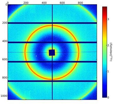
Small-angle X-ray scattering of a micro-vesicle
sample (multilamellar liposomes) using the vacuum-compatible Pilatus
detector, image recorded at a photon energy of 3 keV. The scattering
pattern allows the dimensions of the nano-objects in the examined
sample to be determined. (Source: PTB)
Figure: Small-angle X-ray scattering of a micro-vesicle sample (multilamellar liposomes) using the vacuum-compatible Pilatus detector, image recorded at a photon energy of 3 keV. The scattering pattern allows the dimensions of the nano-objects in the examined sample to be determined. (Fig.: PTB)
For a few months, the new Pilatus X-ray detector has been used for some of PTB's own research projects. At the synchrotron radiation source BESSY II in Berlin-Adlershof, where PTB has been operating its own laboratory for 15 years.
Scientists are now using the new detector, for example, to establish the urgently needed metrological basis for the size determination of microvesicles. A project carried out within the scope of the European Metrology Research Programme (EMRP) and with the significant participation of the Amsterdam Medical Center in the Netherlands is to contribute decisively to fully exploiting the potential of microvesicles for the early diagnosis of diseases.