First implant of electronic eye in Australia
4 September 2012
Researchers at Bionic Vision Australia have successfully performed an implant of an early prototype electronic eye at the Royal Victorian Eye and Ear Hospital in Melbourne.
The bionic eye was implanted in a woman who has profound vision loss due to retinitis pigmentosa, an inherited condition. Ms Dianne Ashworth received the ‘pre-bionic eye’ implant which was switched on last month at the Bionics Institute.
Ms Ashworth reported experiencing some vision: “I didn’t know what to expect, but all of a sudden, I could see a little flash… it was amazing. Every time there was stimulation there was a different shape that appeared in front of my eye,” she said.
The prototype consists of a retinal implant with 24 electrodes. A small lead wire extends from the back of the eye to a connector behind the ear. An external system is connected to this unit in the laboratory, allowing researchers to stimulate the implant in a controlled manner in order to study the flashes of light.
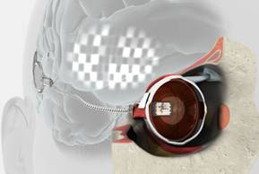
Early bionic eye prototype - electrode array
(image courtesy of
Bionics Institute)
Feedback from Ms Ashworth will allow researchers to develop a vision processor so that images can be built using flashes of light. This early prototype does not incorporate an external camera, yet. This is planned for the next stage of development and testing.
Director of Bionic Vision Australia, Professor Anthony Burkitt, said: “This outcome is a strong example of what a multi-disciplinary research team can achieve. Funding from the Australian Government was critical in reaching this important milestone.”
Sally Capp, Agent General for Victoria in the UK said: “The bionic eye implant is a stunning example of the many innovative medical developments that are being produced by the Parkville Precinct and the wider Victoria medical research community.
Director of the Bionics Institute, Professor Rob Shepherd, led the team in designing, building and testing this early prototype to ensure its safety and efficacy for human implantation. Cochlear technology supported aspects of the project.
“We are working with Ms Ashworth to determine exactly what she sees each time the retina is stimulated using a purpose built laboratory at the Bionics Institute. The team is looking for consistency of shapes, brightness, size and location of flashes to determine how the brain interprets this information. Having this unique information will allow us to maximise our technology as it evolves through 2013 and 2014,” he said.
Although the team claim this is a world first, German company Retina Implant first implanted in human patients in 2005 and started a second clinical trial in 2010. Its multi-centre trial has sites in Germany, the UK, Hungary, Italy and the US. One of the first patients was a teenager in Finland in 2010, and earlier this year patients in the UK and China received implants.
Retina Implant’s subretinal approach to implantation involves placing a 1500-electrode microchip just below the retina, specifically in the macular region. Results of Retina Implant’s first human clinical trial were published in Proceedings of the Royal Society B and showed placement of the implant below the retina, in the macular region, provided optimum visual results allowing patients to recognize foreign objects and to read letters to form words.
Post-implantation, the microchip is turned on — this is when the evaluation of sight restoration begins. As patients must develop new internal processes for interpreting the images they see, it typically takes several weeks to fully realize their new sight capabilities.