Miniature magnetic sensor opens new era for neurology
12 June 2012
A new type of magnetic field sensor, called a chip-scale atomic magnetometer (CSAM), uses miniaturized optics for measuring absorption changes in a rubidium gas cell caused by magnetic fields.
Developed by the US National Institute of Standards and Technology, the sensor has applications in magnetoencephalography (MEG), the study of the brain's magnetic field and the underlying neurology.
The sensor has passed a sensitivity test carried out by Physikalisch-Technische Bundesanstalt (PTB) in a magnetically shielded room. It was able to detect both spontaneous and stimulated magnetic fields of the brain.
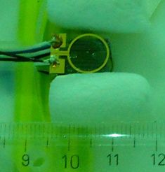
Magnetic sensor the size of a sugar cube
with
electrical and optical lines.
Up to now the measurement of very weak magnetic fields was the domain of cryoelectronic sensors, the so called superconducting quantum interference device (SQUID). They can be considered as the "gold standard" for this application, but they have the disadvantage to operate only at very low temperatures close to absolute zero. This makes them expensive and less versatile compared to CSAMs.
Even though at present CSAMs are still less sensitive compared to SQUIDs, measurements as accurate as SQUIDs, but at lower costs, might eventually become reality. Due to the cooling requirements, SQUIDs have to be kept apart from the human body by a few centimeters. In contrast, CSAMs can be attached close to the human body. This increases the signal amplitude as the magnetic field from currents inside the human body decays rapidly with increasing distance.
Magnetoencephalography (MEG) enables the characterization of neuronal currents, which has gained importance during the last few years for neurologists and neuroscientists. Objective indicators of psychiatric disorders as well as age dependent brain diseases, are urgently needed for the support clinical diagnostics.
Already in 2010 scientists from NIST and PTB had successfully tested the performance of an earlier version of the present CSAM by measurements of the magnetic field of the human heart. For the present study the sensor was positioned about 4 mm away from the head of healthy subjects. At the back of the head, the magnetic fields of alpha waves were detected, a basic brain rhythm which occurs spontaneously during relaxation. In another measurement the brain fields due to the processing of tactile stimuli were identified.
Further information
T. Sander-Thömmes, J. Preusser, R. Mhaskar, J. Kitching, L. Trahms, S. Knappe: Magnetoencephalography with a Chip-Scale Atomic Magnetometer. Biomedical Optics Express Vol. 3 Issue 5, pp.981-990 (2012), http://www.opticsinfobase.org/boe/issue.cfm?volume=3&issue=5
PTB-NIST-Experiment of 2010: S. Knappe, T.H. Sander, O. Kosch, F. Wiekhorst, J. Kitching and L. Trahms. Cross-validation of microfabricated atomic magnetometers with SQUIDs for biomagnetic applications. Applied Physics Letters. 97, 133703 (2010); doi:10.1063/1.3491548. Online publication: Sept. 28, 2010.