Electronic sensor can detect prostate cancer cells
4 June 2012
Scientists at the Barcelona University have developed the main components of a biosensor that can detect even a tiny amount of cancer cells. The sensor uses several hundred nanometric sized optoacoustic biosensors and is dipped into a patient’s urine to detect the cancer cells.
Chances of survival of prostate cancer depend strongly on the stage at which the disease is diagnosed. The new device could improve the diagnosis techniques and avoid fatal treatment delays.
Prostate cancer is the most common malignant tumour and the second leading cause of cancer deaths in men. Men over 50 have a one in five chance of being diagnosed with prostate cancer. An EU research project, called the BOND project, for Bioelectronic Olfactory Neuron Device, is developing a new non-invasive method which could provide an early stage disease detection.
Doctors are also faced with a lack of detailed information about tumours. “Today’s ultrasound examinations are not sufficient to clearly detect the dimensions of a tumour,” explains urologist Stephan Meesen at the hospital in Saarbrücken, Germany. “In practice we see that, unfortunately, we are often wrong or too cautious.” Hence, the need for an integrated biosensor capable of delivering visual information on the progress of tumour cells is essential.
Scientists from the Fraunhofer Institute in St. Ingbert, Germany, have sought inspiration in Greek mythology when naming their imaging biosensor designed to assess tumour dimension. They called their project ADONIS (accurate diagnosis of prostate cancer using optoacoustic detection of biologically functionalized gold nanoparticles).
This project is developing a new sensing technique, relying on optoacoustics. It involves labelling some of the body’s immune defence soldiers, known as antibodies, with gold. The gold-antibody nano-particles construct is put into solution and injected into patients. Thanks to the ability of antigens to bind to specific receptors on the surface of cancer cells, this device delivers gold nano-particles into the tumour tissue.
A laser impulse heats the prostate cancer and induces a pressure wave, which can be detected with an ultra sound probe to assess the shape and size of the tumour. “The gold particles colour the tissue, so this area absorbs more light and sound is produced”, explains physicist Robert Lemor from the Fraunhofer Institute. It is similar to the principle of the radar applied to the inner parts of our body.
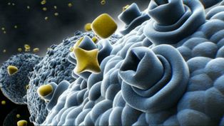
Drawing of gold nanoparticles around cancer
cells
This method generates images based on sending a sound wave and recording the reflection signature after it bounced back from our inner cells. When the tumour is marked with the gold nano-particles, it looks different than the other cells and can be detected. In parallel, the infrared laser beam lights up the tissue and measures its reflection signature. Both together provide the scientists with high sensitive images which can be converted into three dimensional animations.
The basis of this method has already been tested with animals and first studies with humans are in the planning phase. In the future, scientists want to use a higher laser power as a means to heat the gold particles and subsequently destroy the cancer cells.