First 3-D map of invertebrate nervous system
14 Feb 2011
A team of neurobiologists from the University of Freiburg has created the first complete map of all axons that use dopamine as a messenger in a vertebrate, in this case a Zebra fish.
The map identifies all projection possibilities, the so-called “projectome” of every nerve cell for a class of messengers in the nervous system. The research was published in the journal Nature Communications in January [1].
Knowledge of which nerve cells send their connections, called axons, into certain regions of the brain, is particularly important for understanding the functioning of groups of nerves that send out axons to modulate the activity of neural circuits in remote regions of the brain.
One of these groups consists of nerve cells that use the molecule dopamine as a messenger to control many types of behaviour. It is precisely these neurons that die off in people afflicted with Parkinson’s disease, a fact that demonstrates the central role they play in medicine.
The scientists were able to create the first three-dimensional projectome map of the intact brain of the zebrafish by combining the selective genetic marking of individual nerve cells with high resolution microscopy at the ZBSA.
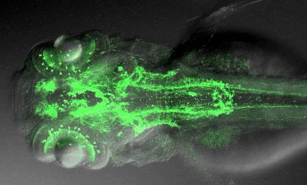
A Zebrafish nervous system.
Source: Driever and Ryu.
The new map reveals important information on the possible functioning of the brain. For instance, it illustrates that dopaminergic neurons of the diencephalon connect distant regions of the brain in previously unimagined ways — regions responsible for higher brain functions in the telencephalon, physiological control in the hypothalamus, the coordination of movement in the hindbrain and the execution of movement in the spinal cord.
These neurons can be involved in effecting changes in basic behavioural states following stress: active reactions like fight or flight or passive reactions like freezing all activity.
In the same study, the scientists describe a new dopaminergic system in another region of the zebrafish’s brain, the corpus striatum, in which the loss of dopaminergic connections in Parkinson patients is particularly severe. The authors speculate that this system might compensate for the low amount of dopaminergic neurons in fish.
In conjunction with further neurobiological studies, the projectome map opens up possibilities for a new understanding of neural circuits in the brains of simple vertebrates like the zebrafish.
Reference
1. Tuan Leng Tay, Olaf Ronneberger, Soojin Ryu, Roland Nitschke, and Wolfgang Driever. Comprehensive catecholaminergic projectome analysis reveals single neuron integration of zebrafish ascending and descending dopaminergic systems. Nature Communications, 25 January 2011. doi: 10.1038/ncomms1171