MRI scans of brain turned into three-dimensional maps of nerves
3 Nov 2010
A team of researchers at the Eindhoven University of Technology has developed a software tool that converts MRI scans of the brain into three-dimensional coloured images of nerve structure. making this visible for the first time without having to operate (video demonstration below).
To know accurately where the main nerve bundles in the brain are located is of immense importance for neurosurgeons, explains Bart ter Haar Romenij, Professor of Biomedical Image Analysis, at the Department of Biomedical Engineering.
As an example he cites deep brain stimulation, with which vibration seizures in patients with Parkinson’s disease can be suppressed: "With this new tool, you can determine exactly where to place the stimulation electrode in the brain. The guiding map has been improved: because we now see the roads on the map, we know better where to stick the needle."
The technique may also yield many new insights into neurological and psychiatric disorders. And it is important for brain surgeons to know in advance where the critical nerve bundles are, to avoid damaging them.
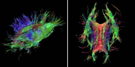
Images of brain 'wiring' made with the tool
developed at TU Eindhoven
The accuracy of the tool is a great step forward. Especially intersections of nerve bundles were difficult to identify till now. Ter Haar Romenij said, "You can now see for the first time the spaghetti-like structures and their connections. But you cannot, of course, dissect a live patient into slices for under a microscope."
The tool was developed by researcher Anna Vilanova, with her PhD students Vesna Prčkovska, Tim Peeters and Paulo Rodrigues.
The tool is based on a recently developed technology called HARDI (High Angular Resolution Diffusion Imaging). The research team developed a system for processing, interpretation and interactive visualization of the very complex data.
Bart ter Haar Romenij expects that the tool can be ready at relatively short notice for use in the hospital within a few years, following validation and further improvements in speed of processing. Currently, for a detailed view, a patient needs to be one hour in the scanner, which is too long. The tool, however, is already widely in use by other scientists.
Video showing the features of the software