MRI scans of brain networks could help predict effects of brain injury
1 April 2010
An MRI scanning technique originally developed for study of brain organization could enable better prediction of the effects of strokes and other brain injuries, neurologists at Washington University School of Medicine in St Louis have found.
The technique, known as resting-state functional connectivity MRI (fcMRI), reveals the health of brain networks that let multiple parts of the brain collaborate. Previous studies have shown that damage to the brain networks helps explain why damage to one brain region can cause problems in abilities controlled by another brain region. The results are published in the March 2010 issue of Annals of Neurology.
Now, for the first time, scientists have linked differences in the nature of the harm done to brain networks to changes in the impairment experienced by patients.
"Clinicians who treat brain injury need new markers of brain function that can predict the effects of injury, which helps us determine treatment and assess its effects," says Maurizio Corbetta, MD, the Norman J. Stupp Professor of Neurology and professor of radiology and of neurobiology. "This study shows that FC scans are a potentially useful way to get that kind of information."
During fcMRI scans, subjects are asked to relax and do nothing. The scans allow scientists to track changes in blood flow to different regions of the brain. During periods of mental inactivity, blood flow in brain regions that are networked generally tends to rise and fall in sync.
In 23 patients who had recently suffered a stroke, researchers looked at two networks: one that directs attention to visual stimuli, and a separate network that controls arm movements. The two networks include regions in both brain hemispheres.
They found patients with damage that disrupted network connections between regions on different sides of the brain had greater functional impairments than patients with damaged connections between regions on the same side of the brain. That meant, for example, that stroke damage on the left side of the brain might lead to problems with control of the right arm, but the losses were significantly worse if the left-side damage disrupted network connections with the right side of the brain.
Those results do not mesh with the traditional picture of brain function, which puts the left side of the brain in control of the right side of the body and vice versa. But this and other recent findings have neuroscientists thinking they may need to adjust that picture.
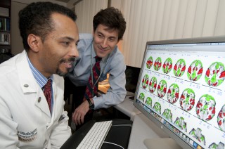
Alex Carter and Maurizio Corbetta have shown
that a scanning approach originally developed for basic studies of
brain organization can also yield useful insights for clinical
treatment of brain injury patients.
"It's not wrong to say that one side of your brain controls the opposite side of your body, but we're starting to realize that it oversimplifies things," says first author Alex Carter, MD, PhD, assistant professor of neurology. "It's starting to seem like proper function requires the two hemispheres to be competing for attention, pushing against each other and thereby achieving some kind of balance."
To further define and confirm FC's clinical applicability, researchers are planning additional studies of brain injury patients. They also plan to use fcMRI in long-term studies of recuperation of such patients.
Reference
Carter AR, Astafiev SV, Lang CE, Connor LT, Rengachary J, Strube MJ, Pope DLW, Shulman GL, Corbetta M. Resting interhemispheric functional magnetic resonance imaging connectivity predicts performance after stroke. Annals of Neurology, March 2010.