Breakthrough in lab-on-chip for fast cancer detection and therapy
14 October 2009
The EU-funded MASCOT project has achieved a major milestone in the development of a lab-on-chip system for the detection of breast cancer.
This is the first time that a lab-on-chip system including many complex sample preparation steps and multiplexed detection has been conceived and is being implemented. All the modules of the diagnostics system for sample preprocessing and detection are ready for further miniaturization and integration in a single lab-on-chip platform. The system will be clinically validated in a breast cancer therapy study in Oslo.
The project is being conducted by IMEC, a leading European research centre in nanotechnology, the Institüt für Mikrotechnik Mainz (IMM), one of the leading European research centers in microfluidics, and their partners in Germany, Norway, The Netherlands and Spain.
Circulating tumour diagnostics
Circulating tumour diagnostics is a promising methodology to individually follow up cancer patients in an early or advanced phase during therapy, thereby improving the doctor's decisions for selecting appropriate therapy.
In the case of breast cancer, 5 ml of blood contains only 2 to 3 tumour cells, so to detect cancer from blood, these rare circulating tumour cells need to be isolated, enriched and their genetic content has to be identified.
Current diagnostics performed in medical laboratories are labour intensive, expensive and time-consuming. They require many sample preprocessing steps in different medical instruments so that the full analysis takes more than a day.
The lab-on-chip system
A lab-on-chip system however can bring huge advantages both to the patient and the healthcare system. They enable a fast, easy-to-use, cost-effective test method which can be performed at regular times in a doctor’s office or even near the patient’s bed. Lab-on-chip systems are a labour-saving and minimally invasive solution for cancer cell detection, therapy selection and monitoring.
The project partners developed a modular platform where each module has its specific task and autonomy and as such can also be used for many different medical applications.
The first module is the incubation module performing the mixing of the blood sample with functionalized magnetic beads which specifically bind the tumour cells. The second module is used for tumour cell isolation and counting using a combination of dielectrophoresis and magnetic sensing with single cell sensitivity.
In the third module, the amplification module, the cell wall of the tumour cells is destroyed and the genetic material (ie the mRNA) is extracted and amplified based on multiplex ligation dependent probe amplification (MLPA).
Within this module, specific assays amplify about 20 markers that are expressed in breast carcinoma cells. In the final detection module, the amplified genetic material is detected using an array of electrochemical sensors. The different building blocks have been developed and validated on spiked blood samples.
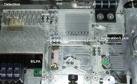
The MASCOT lab-on-chip system.
Photo copyright: MASCOT
The modules are now ready for further hetero-integration into a single lab-on-chip. By miniaturizing and merging the microfluidic and electronic functionalities the reliability and accuracy of the patient’s analysis will be improved. The clinical use of the system will be evaluated to compare it to more conventional approaches in a breast cancer therapy follow-up study.
Within the framework of the MASCOT project, IMEC collaborates with the Institut für Mikrotechnik Mainz (Germany), AdnaGenAG (Germany), Universitat Rovira i Virgili in Sweden, NorwegianRadium Hospital (Norway), MRC Holland (The Netherlands), and FuijerebioDiagnosticsAB (Spain).
The project’s aim is to develop an integrated microsystem for the magnetic isolation and analysis of single circulating tumor cells for oncology diagnostics and therapy follow-up. MASCOT was partly funded by the European Commission (IST-027652).
Bookmark this page