Dual microscope cameras simplify capture of rapid cellular event
31 March 2008
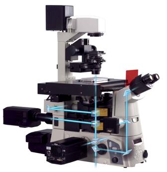
Nikon’s new Ti Series of inverted microscopes can capture images at two wavelengths simultaneously using dual cameras, making it simpler to observe dynamic cellular events.
The ground-breaking stratum structure, which fully utilises Nikon’s 200mm infinity space, makes this possible by allowing an extra camera to be added via the optional back port, in conjunction with a camera on the side imaging port.
The rear camera can be individually centred and focused to guarantee perfect registration of both images simultaneously with no pixel shift. With two cameras it’s possible to accurately observe rapid calcium reactions with dual emission ratiometric dyes such as Indo-1 without dividing the CCD chip and compromising image resolution.
In addition, a dual camera configuration also permits researchers to compare high S/N ratio images when observing interactions between fluorescence proteins with FRET. Each FRET channel can be separated by wavelength and sent to separate cameras. This enables the capture of high-resolution images in the entire frame for each wavelength. Even when the intensity difference between signal intensities is large, a high-quality FRET image can be captured by adjusting camera sensitivity for each wavelength.
Extra components, such as laser tweezers and a photo activation unit, can also be mounted simultaneously on upper and lower tiers of the new microscope.
Making full use of infinity optics, the Ti’s flexible stratum structure allows these components to be used in conjunction with Nikon’s Perfect Focus System (PFS), which is embedded into the Ti nosepiece.
A two-tier epi-fluorescent filter turret holding up to a total of 10 filter cubes can also be incorporated, ideal for multi-user facilities and researchers working with a number of different dyes and fluorescent proteins. Furthermore, for maximum speed and versatility, each motorised filter wheel can be controlled separately.
Commenting on the benefits of the Ti Series’ flexible design, Alan Monk, Biological Imaging Systems Specialist for Nikon Instruments UK said, “The revolutionary design of this system gives researchers working with live cell imaging far greater flexibility in the design of their experimental protocols in fields as varied as neuroscience and embryology.”