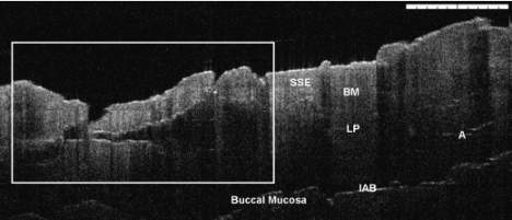
Michelson Diagnostics announces successful OCT imaging of oral cancer tissue25 January 2008 UK optical imaging company Michelson Diagnostics Ltd (MDL) has announced successful initial results from clinical testing, on oral cancer tissue, of its groundbreaking optical coherence tomography (OCT) imaging technology by partner University College Hospital, London (UCH). The tests, performed by UCH scientists on cancerous and suspected cancerous human oral issue, were designed to establish the potential for MDL’s optical imaging technology to revolutionise head and neck cancer diagnosis and treatment. The tests involved comparing images taken by the MDL scanner of the tissue samples, with histopathology images analyzed by trained pathologists. "We are very excited about the breakthrough in image quality that this system offers," said Mr Colin Hopper, Senior Maxillofacial Surgeon at UCH. "OCT could revolutionise the surveillance of pre-cancers in the mouth and eliminate the waiting time for biopsy results. It should also minimise surgery through improved disease mapping. This will provide cost- effective treatments with improved cure rates.” In the tests, 25 oral tissue samples were collected from 14 patients of the UCH National Medical Laser Centre. The samples were scanned with the MDL OCT imaging system and then prepared for analysis by Pathologist Dr Brendan Conn. Clinical Research Fellow Mr Waseem Jerjes said, “The histopathology results showed that it is possible to identify surface structures such as keratin and epithelial layers, the epidermal–dermal junction and areas of cellular crowding, as well as any pathological changes that occur at that level; this is very exciting, this method seems to hold great promises for early in vivo tumour diagnosis.” The company’s optical probe technology provides sub-surface OCT images for research applications in cancer surgery guidance, surveillance and diagnosis.
|