|
|
|
| Diagnostic imaging, neurology | |
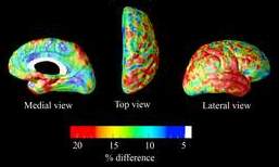
3D MRI imaging highlights brain atrophy in Alzheimer's patients30 October 2007 Researchers at UCLA have used an advanced 3D mapping technique to analyse magnetic resonance imaging (MRI) data to highlight the differences in brain atrophy between mild cognitive impairment and Alzheimer's disease.
The research team analyzed magnetic resonance imaging data from 24 patients with amnestic mild cognitive impairment (MCI) and 25 others with mild Alzheimer’s disease. Patients in both categories exhibit progressive brain atrophy, with most MCI patients showing the pathologic changes characteristic of Alzheimer’s. MCI patients slip into dementia at a rate of 10-15% each year. The research team found that patients with mild Alzheimer’s had 10-20% more atrophy in most cortical areas than MCI patients. The research showed the striking
differences in cortical damage between amnestic MCI and mild Alzheimer’s,
and demonstrated that this innovative three-dimensional mapping technique
greatly outperforms other popular 3D imaging techniques such as voxel-based
morphometry Reference Liana G Apostolova; Calen A Steiner; Gohar G Akopyan; Rebecca A Dutton; Kiralee M Hayashi; Arthur W Toga; Jeffrey L Cummings; Paul M Thompson. Three-Dimensional Gray Matter Atrophy Mapping in Mild Cognitive Impairment and Mild Alzheimer Disease. Arch Neurol. 2007;64:1489-95. |