|
|
||||
| Diagnostic imaging, oncology, surgery | ||||
Successful trial of optical imaging of tissue during cancer surgery13 September 2007 Optical imaging company Michelson Diagnostics Ltd (MDL) has announced successful initial results from clinical testing in cancer surgery of its groundbreaking optical coherence tomography (OCT) imaging technology.
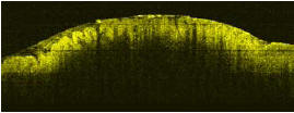
The tests, performed on cancerous and precancerous human oesophagus and lymph node tissue, were designed to establish the potential for MDL’s optical imaging technology to revolutionise cancer surgery. Surgical removal of a tumour is often essential for a cure. Surgeons must be sure that they have cut a safe ‘margin’ of healthy tissue around the tumour to ensure that none is left behind. This is difficult, because removing too much may severely affect the patient’s quality of life and removing too little means that cancerous tissue may be left behind. Surgeons sometimes take biopsy samples of tissue in the tumour margin for laboratory analysis, but have to wait for the result before they can resume the operation. This is very expensive and inefficient. Now MDL offers a potential solution by providing high-resolution sub-surface images of excised tissue during the operation using OCT. The surgeon can scan the tumour margins and decide, in real time, where to cut. OCT provides images of soft tissue at far higher resolution than is possible with ultrasound or MRI scans. The tests, which were conducted by partner Gloucestershire Hospitals NHS Foundation Trust, involved comparing images taken by the OCT scanner of the tissue samples, with histopathology images analyzed by trained pathologists.
Biophotonics expert at Gloucestershire Royal Hospital, Mr Florian Bazant-Hegemark, said “Clinical features of oesophagus tissue and of lymph nodes can be established, in real time, with the MDL OCT scanner. This is very exciting, because it means that OCT scanning has a realistic chance of guiding biopsy and of reducing the need for biopsy, which could speed up cancer operations, reduce the pressure on overloaded pathology departments, and improve outcomes from cancer surgery”. He adds: “The next stage is to confirm these preliminary results in large double-blinded trials”.
|