| Laboratory systems | |||
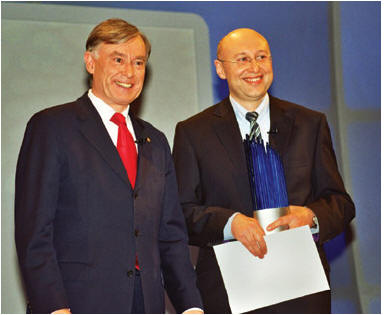
German Future Award for inventor of ground-breaking STED microscope11 December 2006 Mannheim and Wetzlar, Germany. Professor Stefan Hell, Director of the Max Planck Institute for Biophysical Chemistry in Göttingen, was presented with the prestigious German Future Award by Federal President Horst Köhler on 23 November, 2006. This annual technology and innovation award, which was conferred for the tenth time this year, has a cash value of €250,000 and is given in recognition of projects that not only have revolutionary implications for science but are also ready for application and marketing.
Professor Hell received the Future Award for inventing STED microscopy. (STED stands for “stimulated emission depletion”.) The patented STED technique has been licensed to Leica Microsystems, which is developing STED microscopy into a user-friendly instrument to be launched on the market in 2007. The new fluorescence system will be produced in Wetzlar and Mannheim. The Stefan Hell/Leica Microsystems team was already successful at the beginning of the year, when Leica Microsystems won the 2005 Innovation Award of German Industry for the Leica TCS 4PI. This ultra-high resolution microscope system was another invention of Hell’s to be marketed by Leica. Professor Hell was the first to find a way of overcoming the 130-year-old Abbé limit in the fluorescence microscope, the most important microscope in biomedical research. Ever since the 17th century, the light microscope has been one of the main symbols of scientific progress — particularly in biology and medicine. Harnessing STED microscopy, molecules can now be imaged with far greater definition than before. What is new about this technique is the fact that the resolution of microscope images is no longer limited by the wavelength of the light, as postulated by Abbé. The attainable resolution is purely a question of technical design. Hell and his team have already achieved resolutions of 20 nanometers, ie 10 times over the Abbé limit. As protein complexes are in the 10-200 nanometer range, this microscope has the potential to probe life on a molecular scale soon and obtain more accurate information on diseases. The ability to view life in nanometer dimensions opens the door to an understanding of intracellular life processes which was never thought possible before and which may lead to revolutionary discoveries on the subject of how diseases originate. “We therefore expect all well-known universities and research institutes that do basic biomedical research to buy one of these systems in the next few years,” said Dr. Martin Haase, Manager of Leica Microsystems CMS GmbH.
|