| Diagnostic imaging | |
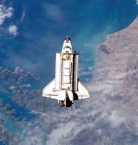
NASA uses live 3D ultrasound imaging to study Space Shuttle astronaut heart mass loss25 September 2006 Andover, Mass., USA. The US National Aeronautics and Space Administration (NASA) is using a Philips ultrasound system to evaluate the effects of space flight on the hearts of Space Shuttle astronauts and, in the near future, astronauts on the International Space Station and ground-based analogs. Of interest to NASA researchers is the loss of heart mass brought on by space flight.
The new technology being used captures a full-volume image of the beating heart in less than a minute and allows physicians to examine the heart as if they were holding it in their hands. It also allows the researchers to make accurate measurements of heart mass, ejection fraction, blood flow, strain rate and cardiac wall motion pre- and post-flight. "We have a very short window of time in which to do an echo exam on the astronauts," said David S. Martin of Wyle Laboratories, Inc., ultrasound lead for the NASA Cardiovascular Laboratory at the Johnson Space Center in Houston, Texas. "Live 3D Echo allows us to quickly grab all the image data we need to do a full examination of the heart anatomy and function and send the astronauts on their way. Following the image acquisition, we use off-line analysis software to do several measurements that help us evaluate changes after space travel." The use of this heart imaging and measurement technology will be part of ongoing research at the NASA Cardiovascular Laboratory. It will also complement the imaging done by a modified Philips HDI 5000 ultrasound system that was installed in the International Space Station's Human Research Facility in 2001. "These new ultrasound technologies help us efficiently conduct sophisticated cardiac research of astronauts and the effects of microgravity," said Martin.
|
 Royal
Philips Electronics (NYSE: PHG, AEX: PHI) has announced that NASA will use
its iE33 echocardiography system and QLAB quantification software to monitor
the astronauts. Launched in 2004, the iE33 is a new generation of cardiac
ultrasound equipment that uses high-definition imaging to help physicians
diagnose heart disease and incorporates data-analysis tools to help support
treatment decisions and monitor treatment success.
Royal
Philips Electronics (NYSE: PHG, AEX: PHI) has announced that NASA will use
its iE33 echocardiography system and QLAB quantification software to monitor
the astronauts. Launched in 2004, the iE33 is a new generation of cardiac
ultrasound equipment that uses high-definition imaging to help physicians
diagnose heart disease and incorporates data-analysis tools to help support
treatment decisions and monitor treatment success.