| Diagnostic imaging, oncology | |||
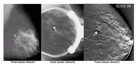
More sensitive imaging system for detecting breast cancer12 December 2005 Duke University Medical Centre and Siemens Medical Solutions are collaborating to develop a digital breast tomosynthesis (tomo) system that could detect mass lesions often undetectable by conventional mammography. The new system is based on Siemens’ Mammomat NovationDR full-field digital mammography system. A prototype was presented at the recent Radiological Society of North America (RSNA) congress in Chicago. The new prototype employs a Mammomat NovationDR system that has been modified with a motorised X-ray tube to acquire image projections of the breast from many different angles, as well as a digital detector for increased speed and quality image capture. Preliminary results conducted at Duke University with several mastectomy and patient cases suggest that the new tomo system is able to detect subtle mass lesions otherwise difficult to pick up with standard mammography. When fully developed, this new solution is expected to further enhance diagnostic ability by improving sensitivity of breast cancer detection and more importantly, increase workflow by reducing false positives/misdiagnosis and unnecessary biopsies. Holger Schmidt, President, Special Systems Division, Siemens Medical Solutions, said: “Through a dedicated team of researchers at both Duke University and Siemens, we have developed a breast tomosynthesis system. We hope it will be a major step in diagnosis that will revolutionise the way breast cancer is treated.”
“Breast tomosynthesis may be one of the first techniques that can actually replace conventional 2D mammography,” said Joseph Lo, Ph.D., Assistant Research Professor of Radiology, Duke Advanced Imaging Laboratories, Duke University Medical Centre. “While mammography is sensitive at finding cancer, it sometimes results in false positives — leading to workflow issues for the clinician and unnecessary strain on the patient. The goal of our studies is to reduce these false positives by providing 3D information at the same high resolution and reasonable dose as mammography, while also improving patient comfort.” Featuring a large field-of-view enabled by a large area detector, the prototype system is designed to be suitable for nearly all breast sizes. It has the potential to provide high sensitivity and specificity that could allow radiologists to detect and characterise suspicious lesions more precisely. With low-dose 3D mammography it may eliminate normal tissue overlap, which might otherwise obscure lesions. Additionally, enhanced visibility to view masses may be made possible with high contrast resolution. "If digital breast tomosynthesis fulfills its promise of providing the unusual combination of greater sensitivity coupled with greater specificity and improved patient comfort, it may rapidly replace routine 2D mammography in the screening setting,” said Jay Baker, M.D., Division of Breast Imaging, Department of Radiology, Duke University Medical Centre. “While careful clinical trials are required to confirm the potential advantages of tomosynthesis, initial testing has been encouraging and tomosynthesis images have demonstrated extraordinary anatomic clarity." Ongoing studies are underway in an effort to improve upon the existing prototype and to optimise radiographic techniques to produce superb image quality with reduced dose. Researchers are also exploring a new acquisition-and-viewing workstation based on Siemens’ syngo platform with the intent to enhance image processing and workflow. Preliminary clinical trials are planned to begin in the coming months.
|