| Diagnostic imaging | |
Functional MRI is new tool in early diagnosis of bronchial asthma18 August 2005
One out of ten children as well as every fifth adult in Germany suffers from bronchial asthma — and this number is rising. The density of lung tissue plays a special role with asthmatics. During an asthma attack the muscles of the bronchial tree become tight, reducing air flow out of the lungs, that is, part of the air remains in the lungs. For asthmatics the density of the lung tissue increases only partially or not at all during exhalation. A wide range of treatment options is available if the disease is diagnosed early and correctly. To date, lung function measurements have been used to measure airflow into and out of the lungs as well as the velocity of flow. The new technique allows experts to determine which part of the lungs is involved. “Using functional MR of the lungs, the treating physician is able to not only detect the areals of the lungs involved but also at what points in time the damaging air remains in the lungs,“ explained Thomas Rupprecht, M.D., team manager of the clinical trial, and chief physician at the Clinic for Children and Adolescents in Bayreuth as well as Visiting Professor at the University of Erlangen. Conventional lung function measurements can only determine if the patient is suffering from bronchial asthma, not which parts of the lungs are affected.
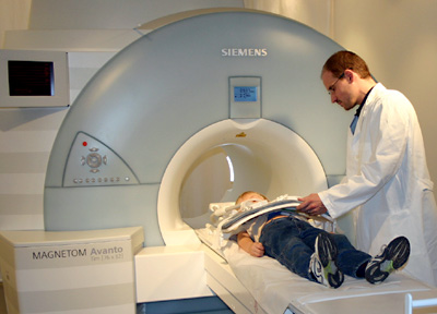
Photo: Chief Physician Dr. Hans-Georg Topf prepares a
child for preventive At a young age, the performance ratio of a healthy person’s lungs is much higher than actually required — this means that a child suffering from bronchial asthma lives without side effects and performs as well as its classmates. Over time, however, the child’s performance decreases rapidly and the disease becomes evident. Conventional methods make it especially difficult to diagnose the illness at an early age. Essentially, the methods only measure the lung volume which is minimally different for sick or healthy children. Yet it is important to obtain precise information about the density of lung tissue for children and adolescents. The new method measures the actual ventilation of the lungs as well as precisely locates pathological areals. It allows for diagnosing and treating bronchial asthma before a decrease in performance confirms the disease.
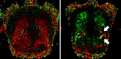
Image: An MR image showing a healthy lung on the left, on the right a lung of a patient who suffers from bronchial asthma: The dark places mark the areals with no respiratory exchange. Thus the physician is able to detect diseased places of the lung exactly. The green and red areals of both images stand for the respiration process. The green demonstrates the inhalation and the red exhalation. The basis for this type of measurement is the examination of the patient with magnetic resonance. In the course of several breaths, the MR system acquires images inside the lungs and loads them into the newly developed software which analyzes each cubic centimeter of the density plot. It is highly suitable for clinical routine as proven by the study involving over 100 patients of the University Clinic Erlangen-Nuremberg. Bronchial asthma is a chronic inflammation and oversensitivity of the bronchial tubes which leads to periodic attacks of shortage of breath, coughing, and chest tightness. The cause is a pathological response of the lining of the air passages to different stimuli. When the pathology is already diagnosed in infancy, early therapeutic measures provide a cure or a reduction in symptoms. Source: Siemens Ag |
 A
new method for detailed diagnosis of bronchial asthma using functional
magnetic resonance imaging (fMRI or fMR) has been developed by the
University Clinic in Erlangen, Germany, together with Siemens Corporate
Research (SCR) in Princeton/USA.
A
new method for detailed diagnosis of bronchial asthma using functional
magnetic resonance imaging (fMRI or fMR) has been developed by the
University Clinic in Erlangen, Germany, together with Siemens Corporate
Research (SCR) in Princeton/USA.