| Diagnostic imaging | |
European premier for combined single photon emission CT and diagnostic CT at Erlangen Hospital15 April 2005 The Nuclear Medicine Clinic and the Radiological Institute at Erlangen University Hospital have begun using Europe’s first combined system for SPECT (Single Photon Emission Computed Tomography) and diagnostic Computed Tomography from Siemens Medical Solutions, the Symbia TruePoint SPECT•CT. Erlangen University Hospital is one of only two institutes worldwide to clinically implement this new SPECT•CT procedure. This opens new opportunities for physicians in detecting tumours and cardiac diseases: diagnosis with the new system is earlier, more precise, and more reliable. This in turn significantly improves the patient’s chance of being cured.
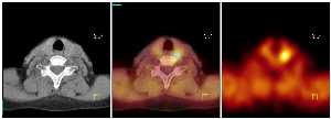
Symbia is the first to combine the functional sensitivity of a SPECT system with the detailed anatomical information provided by diagnostic multi-slice CT systems. SPECT, the nuclear medicine imaging procedure, enables organ function, cell metabolism, and other functional parameters in the human body to be displayed. Metabolic processes are displayed using different radioactive substances given to the patient in extremely small doses. As a result, pathological changes can be detected at the molecular level before changes in the anatomical structure at the submillimeter level are visible with CT. Due to the high specificity of the radioactive test substances used however, the detailed anatomical information that is also required is limited, and precise localization of the diagnostic findings is often difficult. Nuclear medicine examinations are function oriented, as opposed to structure-oriented procedures such as computed tomography. Combining nuclear medicine technology with a Spiral CT brings together the advantages of both procedures and significantly increases the diagnostic precision of SPECT. After the examination, the CT slice images are superimposed on the SPECT images, enabling the physician to detect the location of diseases in the body with submillimeter precision. This new technology will be used at Erlangen University Hospital in particular for diagnosing tumors and cardiac diseases. For example, additional metastases will be detected in patients with thyroid cancer using the highly-sensitive SPECT procedure. If the tumour has spread, CT images generated with the hybrid system can be used to localize the metastases in the body. After the examination with SPECT•CT, the physician can determine whether treatment should continue depending on the results. When searching for bone metastases, the combination of a skeletal scintigram and CT increases diagnostic precision. For the patient, this also reduces the period of uncertainty waiting for a definitive diagnosis. After a heart attack, the system enables fast and precise determination of the location and degree of damage to the heart muscle due to insufficient blood supply. Improved therapy planning optimized for each individual patient reduces unnecessary operations and lowers risk when surgery is required. “Today, nuclear medicine is the most powerful molecular imaging technique for non-invasive metabolic examination of the patient. The combination with diagnostic CT significantly improves all aspects of SPECT imaging. In particular, it compensates for SPECT’s limited spatial resolution capabilities,” according to Professor Torsten Kuwert, Director of the Nuclear Medicine Clinic at Erlangen University Hospital. In Cardiology, TruePoint SPECT•CT offers valuable information regarding cardiac function and blood circulation. The new technology uses diagnostic multi-slice CT to calculate the attenuation correction and provides a precise display of attenuation data, which is reflected in increased specificity and more reliable findings when diagnosing cardiac blood circulation problems. “The introduction of TruePoint SPECT•CT and the Symbia product family underscores Siemens deep commitment to developing modern technology as an answer to the clinical requirements of our partners,” said Dr. Erich R. Reinhardt, CEO of Siemens Medical Solutions. “TruePoint SPECT•CT meets the current and future expectations of nuclear medicine because it maximizes the information at the molecular level in combination with precise anatomical details. With this new technology, our customers, physicians, can precisely localize metastases in their patients’ bodies and analyze them in terms of size, type, and degree.” Background information: Physicians can use the Symbia platform with TruePoint SPECT•CT technology
to perform three separate types of examination: SPECT, multi-slice CT, and
SPECT•CT, all from a single system. Thanks to its small footprint, Symbia
easily integrates into a variety of clinical environments. Symbia offers
various multi-slice CT configurations, with rotation speeds of up to 0.6
seconds per rotation. This produces high-quality CT scans in just a few
seconds. As a result, physicians can produce functionally exact and
anatomically precise SPECT•CT studies significantly faster than with any
other system on the market. Nuclear medicine enables physiological and biochemical processes and their pathological changes to be localised and examined in a non-invasive manner. In contrast to other imaging procedures, nuclear medicine examinations are function oriented. As a result, metabolic processes can be examined using various radioactive substances with short half-lives, and displayed in functional images. The substances accumulate in different types of tissue depending on their pharmacological characteristics. During decay, the substances emit gamma radiation, which behaves similarly to X-radiation. As a result for example, tumours can be visualized in very early stages. Or, after a heart attack, it enables fast and precise determination of the location and degree of damage to the heart muscle due to insufficient blood supply.
|