| Diagnostic imaging | |
CT scans unwrap secrets of British Museum's Egyptian mummies7 April 2005
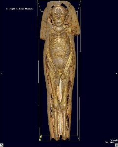
Santa Ana, Calif, USA. In conjunction with its new exhibition, Mummies: Death and the Afterlife in Ancient Egypt, the Bowers Museum has conducted the largest collection of CT scans ever performed on Egyptian mummies. The Bowers Museum, a team of local radiologists and international curators, and GE Healthcare, a unit of General Electric Company (NYSE:GE), announced today the results of computed tomography (CT) scans of six ancient Egyptian mummies from the renowned collections of the British Museum. The mummies are the focus of the Bowers Museum's upcoming landmark exhibition, Mummies: Death and the Afterlife in Ancient Egypt, which opens April 17, 2005. Radiologists discovered that the adult mummy of a Roman man, possibly from Thebes, who lived approximately 140-180 AD, was buried with all of his internal organs. Dr. M. Linda Sutherland, Fellow of the Bowers Museum and Member of the Board of Directors and Partner of Moran, Rowan & Dorsey (MRD), Inc., a diagnostic radiology group in Orange County, Calif., believes this is possible evidence of early embalming practices. "Based on the CT scans we performed, we believe this male was embalmed with some type of fluid containing a metal," said Sutherland. "In the images we were able to see high-density spots in his brain, liver and muscles. I believe this new information will help challenge the conventional wisdom of ancient embalming techniques." The scans performed at the Bowers Museum represent the largest collection of CT scans ever performed on Egyptian mummies. Researchers plan to use the images to determine how these people lived, their age, their overall health, cause of death, and how they preserved their bodies. The whole-body scans of the six mummies will continue to be reviewed and interpreted by MRD Inc. radiologists and British Museum curators. CT scans and X-rays have been performed on mummies in the past, yet none have employed the advanced technology that was utilized at the Bowers Museum, according to Bowers Museum President Peter Keller. "At last, the public has had a glimpse into what lies beneath the linen wrappings of these mummies and the ancient practices that have preserved these human remains through the centuries," said Keller. CT scanning and virtual reality imaging involves acquiring a number of cross-sectional images, or "slices" of the body. Those images are then fed into a workstation that enables radiologists and researchers to see the images in 3-D. "GE's state-of-the-art CT equipment is enabling noninvasive, high-resolution, three-dimensional views of these ancient treasures," said Gene Saragnese, Vice President and General Manager of the GE's Global Functional and Computed Tomography (FCT) business. "GE is proud to donate the use of our equipment to facilitate this historic event, which is enabling researchers to shed new light on the life and times of our ancient ancestors." The mummies scanned by GE's LightSpeed CT include:
See also Results of Tutankhamen scan revealed CT scans and DNA tests help unveil mystery of long-lost female pharaoh |