Biomagnetic separation attracts diagnostics, DNA transfer and cancer therapy research
27 May 2011
A brief history of biomagnetic separation
The principle behind magnetophoresis is the action of a magnetic force on particles. Even a simple block magnet can exert some degree of force onto a nearby test-tube. The process of refining this with improved orientation of the magnets and fluid (which is to be separated) and numerical algorithms has lead to SEPMAG, a specialized company, being formed and producing commercial magnetic separation products with a wide range of applications within the industry.
In becoming the successful and promising tool that biomagnetic separation is now widely seen as, a number of obstacles had to be overcome. The maximum capacity manufactured by IVD companies using the previous generation of biomagnetic separators was around 50ml: well below the several litres often required for manufacturing processes and the widely used Chemoluminescence Immunoassays (CLIA).
Further concerns were raised by the safety of these machines, since there had been reported injuries and the problems caused by having to remove pacemakers, computers or magnetic recording devices from around a large surrounding area, because of possible magnetic interference. These obstacles presented significant challenges to the development of bio-magnetic separation, such that non-magnetic separation research became increasingly popular in the hope of providing a realistic alternative.
These challenges were addressed in 2004, through discussions generated by the collaboration between ATIPIC and BIOKIT; and resulted in successful and intrinsically safe scaling up of the process and a reduced separation time (to one order of magnitude).
This impressive work was further built upon by subsequent dialogues between a number of global-reaching IVD companies such as Bio-Rad, Biocode, Diasorin and Roche Diagnostics. The major change resulting from this was in the definition of the bio-magnetic separation process, since analysis of the process was now available using a range of parameters.
Defining the operating conditions in addition to separation time provided additional support for developing SOP (standard operating procedure) and Quality Assurance protocols, which are both important milestones in a products journey to the market place.
Background to SEPMAG
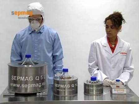
The potential of this technique within the biotechnology and biomedical industry is demonstrated by the decision taken by Dr Luis M. Martinez to purchase the patent of the product resulting from international discussions in 2007 and form a company specialising in this area, SEPMAG.
The devices that SEPMAG subsequently produced enabled the study of the dynamics of colloidal suspension, thanks to the homogeneous conditions provided by this new generation of bio-magnetic separation devices. Another consequence of this technology was the generation of new theoretical magnetic colloidal models to explain the greatly reduced separation times seen experimentally with the SEPMAG devices.
SEPMAG was in a position to capitalise on the numerous benefits of bio-magnetic separation, and in early 2011, secured a Chinese Patent for its unique homogenous biomagnetic separation process from China’s State Intellectual Property Office (SIPO). They have since entered the Chinese market and shipping of the SEPMAG Q250ml product to CapitalBio began in January. CapitalBio is a leading Chinese company, which has recently begun a partnership with Roche Diagnostics. Because of the advantages of bio-magnetic separation over the market alternative, ELISA (Enzyme-Linked ImmunoSorbent Assays), it is hoped that acquiring this strategically significant client is part of a growing trend in the uptake of SEPMAG’s products in both the Chinese and global markets.
Josep Maria Simó, CEO of SEPMAG, commented: “We are delighted that the Chinese Patent Office has granted SEPMAG a patent for our unique magnetophoresis techniques. This will allow us to further consolidate our Far-East presence, having already established a strong and growing foothold in Japan. Over the next couple of years, we forecast the Chinese marketplace to be amongst the fastest growing for bio-magnetic separation systems. The agreement we have signed with CapitalBio marks the start of a significant expansion in the region and a continuation of the internationalisation of SEPMAG technologies.”
To bring the advantages of bio-magnetic specification to the wider biotechnology industry, SEPMAG has been working with theoretical physicists developing ‘precision magnetophoresis’ to enable the application of the technology to a wide range of specific uses across the industry.
Current uses of BMS
Those benefiting today from these developments in the field of
BMS are from a broad range of specialities within the biotechnology
industry. They do however fall into two rough categories; those
using it for in vitro procedures and those applying it to
in vivo scenarios.
The in vitro applications
fall into 4 categories [1], all of which are in frequent use across
the industry:
- Immuno and molecular diagnostics: Bio-magnetic separation increased [2, 3, 4]) or DNA can be captured and separated from a sample. The process is also easy to automate and as a result some diagnostic companies have developed their own analysers for the process.
- Protein purification: Capture of specific proteins can be achieved in the face of high exposure levels and the presence of particulates. Immobilisation of beads with magnetic forces makes it possible to concentrate and purify the selected protein [6]. Because the filtration, centrifugation and clarification stages of conventional separation are not needed when separating using magnets, more protein can be recovered from the original suspension than is possible using conventional methods. This purification allows for downstream processing, used commonly in pharmaceutical development and production.
- Cell capture: Using crude samples such as
blood, bone marrow, tissue homogenates, stool, cultivation
media, food, water and soil, BMS can capture specific cell types
[7]. There are two possible methods of capturing cells:
- Using high magnetic forces (magnetic columns) with cells that have adequate intrinsic magnetic moment (namely erythrocytes with high concentrations of paramagnetic haemoglobin and magnetotactic bacteria);
- Using permanent magnet devices and labels for all other cell types. These magnetic components are able to interact with the cell surface via high affinity ligands;
- The capture of specific cells then opens the doors for further investigation or diagnosis [8]; for example, cultivation of cells or analysis of intercellular components (by lysis of the cell) — since it is possible to remove the magnetic label if needed.
- Nucleic acids capture: BMS can be used to collect all the nucleic acids (DNA or RNA) in a suspension, using silica magnetic beads. This precedes the separation of specific nucleic acid sequences using magnetic beads attached to the correct complimentary DNA strands. The BMS technique provides an effective alternative to the time-consuming PCR step, thus opening the doors to further DNA research.
The benefits of BMS
The benefits of bio-magnetic separation are six-fold. The systems operate such that there are high degrees of reproducibility for every single diagnostic test (an essential factor in any scientific experiment or test). The scale up of operations is easy, which simplifies any revalidation processes.
Process monitorisation allows any problems (for example bead size distribution, concentrations and buffer conditions) to be detected earlier, which avoids the production of defective batches and increases the efficiency of the system. Safe operation is achieved with minimal stray magnetic fields, with the additional bonus of saving space in the lab by reducing the caution distance of operation to a few centimetres.
The time taken for the separation process is reduced, taking the separation time down from hours to seconds and without the need for excessive force — which generates additional problems. Further gains are made financially by the minimal loss of beads and precious bio-molecules through the process; up to 20% savings have been reported by some companies.
Future and potential applications of BMS
The hardware developed by SEPMAG opens the door to the potential application of this technology in a host of laboratory and clinical settings. A lot of this potential lies in its in vivo application.
Building on the nucleic acid capture already in use, the transfer of nucleic acids (as foreign genetic material) into cells is being researched. Using magnetic forces that act on molecules attached to the nucleic acids directs them towards targeted cells. This can increase the transgene expression levels by up to three orders of magnitude from the normal method, increasing the efficiency of the process. This has potential applications in bacteriology, pharmaceutical R&D and genetic studies.
The conditions for viral-mediated gene delivery (an alternative mechanism of DNA transfer) may be optimised by combining the viral vectors with magnetic particles. This allows the viral dose and exposure to be reduced, which in turn reduces the chance of any side effects.
Magnetic separation technology has further roles in the delivery of biological compounds, including pharmaceutical molecules. If associated with magnetic nanoparticles, therapeutic agents can be targeted to direct sites in the body [9]. This overrides the need for ligand-based drug delivery, the problems of which are twofold. Firstly, specific receptors may not be 100% specific, causing additional side effects and secondly, identifying these receptors can be a challenge, often creating impassable obstacles for conventional drug delivery.
Magnetically-mediated hyperthermia [10] (MMH) is a possible physical therapy for cancer, based on the principles of heating tumour cells to above 43 degrees to kill them. Current hyperthermia-inducing methods are limited by their inability to be localised to an appropriate degree. However, magnetic forces can be used to accumulate magnetic particles in a localised region of tissue.
The accumulated magnetic particles are then subjected to an alternating magnetic field and heat up, killing the surrounding tumour cells. The selectivity of this treatment lies in the fact that cancer cells are less heat-resistant than normal cells. This specificity is associated with reduced side effects and it would then be possible to use this alongside conventional cancer therapy (such as chemotherapy and radiotherapy).
Tissue engineering is a rapidly expanding new dimension of modern medicine, and magnetic-force based tissue engineering [11] — which uses the same magnetic forces of BMS — is at the heart of it. Labelling cells magnetically allows them to be organised into 2D and 3D multi-layered structures using magnetic forces. Studies have already used it for a range of tissues from umbilical veins to retinal pigment epithelial cells and keratinocytes to mesenchymal stem cells. These functional substitutes could one day be used in the clinic to replace lost or damaged tissues.
Conclusion
The parameterisation of the bio-magnetic separation process allows for optimisation of the process, which in turns opens the doors for its application to a wide range of clinical and laboratory processes. In the last decade this technology has improved unrecognisably and has been applied to an enormous range of situations. With the current research on in vivo applications, it is an exciting time for biomagnetic separation and the large number of specialities, companies and patients that look to benefit from it.
References
- Corchero, J. L., Villaverde, A., 2009. Biomedical applications of distally controlled magnetic nanoparticles. Trends in Biotechnology, Vol.27 (No.8).
- Deponte, S., Steingroewer, J., Löser, C., Boschke, E., and Bley, T. 2004. Biomagnetic separation of Escherichia coli by use of anion-exchange beads: measurement and modelling of the kinetics of cell-bead interactions. Analytical and Bioanalytical Chemistry, Vol. 379 (No. 3) 419-426.
- Steingroewr, J., Bley, T., Bergemann, C., and Boschke, E. 2007 Biomagnetic separation of Salmonella Typhimurium with high affine and specific ligand peptides isolated by phage display technique. Journal of Magnetism and Magnetic Materials, Vol 311(1) 295-299.
- Scott, D. L., Clark, C. W., Tooley, P. W., Carras, M. M., Maas, J. L. 2000. The use of Biomagnetic separation to recover DNA suitable for PCR from Claviceps species. Letters in Applied MicroBiology, 31(2) 95-99.
- Demidov, V. V., Bukanov, N. O., and Frank-Kamenetskii M. D., 2009. Duplex DNA Capture. Current Issues in Molecular Biology, 2(1) 31-35.
- Safarik, I., and Safarikova, M. 2004. Magnetic techniques for the isolation and purification of proteins and peptides BioMagnetic Research and Technology, 2(7).
- Safarik, I., and Safarikova, M. 1999. Use of magnetic techniques for the isolation of cells. Journal of Chromatography B, 722, 33-53.
- Olsvik, O., Popovic, T., Skjerve, E., Cudjoe, K. S., Hornes, E., Ugelstad, J., Uhlén, M. 1994. Magnetic separation techniques in diagnostic microbiology. Clinical Microbiology Reviews, 7(1) 43-54.
- Dobson, J. 2006 Magnetic Nanoparticles for Drug Delivery. Drug Development Research (Nanobiotechnology), 67 (1): 55-60.
- Moroz, P., Jones, S. K., Gray, B. N., 2002. Magnetically mediated hyperthermia: current status and future directions. International Journal of Hyperthermia, 18(4) 267-84.
- Ito, A., Ino, K., Hayashida, M., Kobayashi, T., Matsunuma, H., Kagami, H., Ueda, M., Honda, H. 2005. Novel Methodology for Fabrication of Tissue-Engineered Tubular Constructs Using Magnetite Nanoparicles and Magnetic Force. Tissue Engineering, 11 (9-10): 1553-1561.
Source: SEPMAG